For Professionals

Personalized Breast Cancer Screening For Better Detection in Dense Breast Tissue
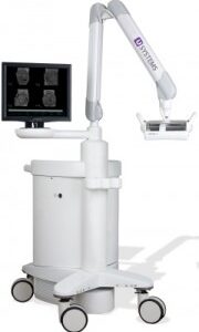
3D Automated breast ultrasound ( ABUSTM) is the latest technological advancement in breast ultrasound imaging from GE Healthcare, specifically designed to address the functional limitations of screening mammography for women with dense breast tissue affecting over 40% of women considered to be at a 4-6 times higher risk for breast cancer. It’s the only FDA and Health Canada approved ultrasound breast cancer screening device indicated for enhanced surveillance as an adjunct to mammography, particularly for ongoing mammographic breast density cases of normal or benign findings. Screening ABUS has been shown to offer clinicians and their patients a life-saving opportunity for the detection of previously undetectable and suspicious cancers in dense tissue that are potentially smaller, less invasive, node negative, and at an earlier more treatable stage requiring less invasive protocols.
ABUS Delivers Exceptional 3D Image Quality for Complete Breast Mapping and Accurate Interval Comparison
ABUS delivers exceptional 3D image quality using a large, automated scanning probe that virtually eliminates operator skill variability and completely maps and triangulates the entire breast area for accurate interval comparison between studies, helping to deliver greater clarity and peace of mind to both patients and healthcare providers. The ABUS scanning probe is gently positioned at three different locations on each breast by a licensed and ABUS-certified sonographer, allowing for the acquisition of approximately three hundred image slices per scan, totaling 900 image slices per breast, from the skin’s surface back to the chest wall. In just minutes, 3D-reconstructed images are rendered using proprietary software, capturing precise anatomical detail of complex breast tissues and structures to be reviewed by the radiologist who can virtually peel back the layers (or image slices) of breast tissue to visualize suspicious abnormalities which may be hidden by dense tissue on a mammogram. Although the sensitivity, specificity and reproducibility of ABUS is proven to be high, it’s accuracy is optimized by the reading radiologist who is trained and experienced in the interpretation of ABUS images.
The complete field of view covers 15cm x 17cm x 5.0cm. The machine has both a high center-frequency enabling high resolution and ultra broadband characteristics for optimal contrast. The standard quality and utility of ABUS is exponentially better than handheld ultrasound which is limited by operator-skill variability, and limitations in acquiring triangulated reference data of the entire breast area for accurate interval comparison. For these reasons handheld is primarily indicated as a valuable followup diagnostic imaging tool for spot-checking suspicious areas of concern identified in a screening mammogram or ABUS.
Helping Clinicians Look Differently at Dense Breast Tissue One ABUS Scan at a Time – Mammogram Alone Is Not Enough
Clinicians are now incorporating ABUS as part of a more personalized and preventative screening strategy for their patients to help find the up to 50% of cancers that mammography potentially misses in dense breasts. Multiple studies have shown that 91% of ABUS-detected cancers were confirmed as invasive, smaller, non-negative and more treatable in various asymptomatic screening populations with higher breast density when used to augment normal or benign mammograms.
ABUS is Not just for Women with Dense Breasts
ABUS is also recommended by many prevention-focused clinicians for general breast cancer screening with an interest in acquiring earlier baseline and comparative information for a timely understanding of subtle tissue changes that can be monitored and proactively addressed as needed, well before breast cancer develops. As supplementary screening information is acquired at an earlier stage using ABUS, patients become much more engaged and comfortable about their own breast health as part of an ongoing learning process which leads to better decision-making, more available and less aggressive treatments, and healthier outcomes.
All women at every stage of life are taking advantage of ABUS regardless of their medical history, including younger, pregnant and lactating women, or other individuals at higher risk (ie. hormone therapy, surgical history [augmentation, reduction, biopsy, lumpectomy, mastectomy, or reconstruction] who don’t easily qualify for OHIP-covered methods but prefer earlier and proactive baseline screening and monitoring. Many patients with dense tissue whose annual or bi-annual mammograms have been consistently normal or benign, are introducing ABUS more frequently into their regular screening protocol for its added value and to help minimize their cumulative radiation risks and tissue discomfort and stress from the heavy compression of mammography.
ABUS Meets Quality Assurance Standards to Protect Patient Care
ABUS is not currently covered under OHIP as part of the Ontario breast screening program (OBSP), however it is currently accessible at the PreView Breast Screening clinic in Barrie whose licensed sonographers, radiologists and support staff, work responsibility to comply with the clinical practice parameters and imaging standards of the Ministry of Health and Long-Term Care for independent health facilities, and with the breast ultrasound examination and teleradiology standards of the Canadian Association of Radiologists.
Clinicians Must Fulfill their Leadership Role in the Breast Screening Process
Through better access to research, healthcare professionals are becoming much more informed about breast density, the limitations of screening mammography, and the evidence-based advantages of ABUS as a valuable adjunctive screening tool. Consequently, a growing number of clinicians and their patients are advocating for breast density reporting and notification from their mammogram reports and screening supplementation with ABUS. As patients become better informed about their breast health, healthcare providers are obligated to meet each patient’s own unique breast health needs and to minimize their risk factors based on reliable data from a more proactive and personalized breast screening program including mammography, adjunctive ABUS, clinical/self breast examinations and supporting lab data. As with 33 out of 50 states in the USA, provincial health ministries in Canada are finally beginning to follow suit, starting with the province of BC which legislated breast density reporting and notification in October 2018.
![]()
![]()
Considered the largest trial ever undertaken by an ultrasound company, the SOMO−INSIGHT was a multi-center nationwide clinical research study in the US, sponsored by U-Systems who worked with some of North America’s leading breast imaging experts to evaluate the ability of somo-v ABUS to improve cancer detection rates for women with greater than fifty percent dense breast tissue. Up to 20,000 patients were enrolled in the study which resulted in a significant number of mammographically negative breast cancers that would not have otherwise been detected had the participants not had an ABUS exam.
![]()
The EASY Clinical Study (European Asymptomatic Screening Study) is a large European clinical research study sponsored by U-Systems Inc. to evaluate the integration of ABUS into the routine practices of a high-volume hospital-based breast cancer screening program in Stockholm, Sweden. The clinical study was designed to determine whether Full Field Digital Mammography combined with ABUS could improve breast cancer detection when compared to mammography alone in women with dense breast tissue.
The study protocol accommodated the standard screening practices in Sweden and outlined a process for integrating ABUS review into an existing double-reading system where two different radiologists independently reviewed breast image exams.
The EASY study recruited 8,000 asymptomatic women with dense breast tissue and although the results of the primary analysis were not intended to reach statistical significance, they yielded valuable trending information about the potential impact on existing cancer detection rates of a multi-modality approach combining ABUS with routine mammography to improve the accuracy of routine screenings for women with dense breast tissue.
![]()
Click here for mammographic breast density and 3D Automated Breast Ultrasound (ABUS) research and general interest articles.
